Anatomy Of Chest Bone - Rotation of 3D skeleton.ribs,chest,anatomy,human,medical ... - It is comprised of many bones, formed by intramembranous ossification, which are joined together by sutures (fibrous joints).
Anatomy Of Chest Bone - Rotation of 3D skeleton.ribs,chest,anatomy,human,medical ... - It is comprised of many bones, formed by intramembranous ossification, which are joined together by sutures (fibrous joints).. The medial anterior chest is defined by the sternum, which consists of 3 flat polygonal bones: Anatomy is the amazing science. Irregular bones have complex shapes. Human chest bone structure parts of the chest bones. When a patient flexes the neck forward, the prominent process is usually that of the 7th cervical.
And we want to know some borders about it. Chest bone, ribs, lung, heart, xiphoid process. Vascular anomalies of aorta, pulmonary and systemic vessels. Atlas of wrist mri anatomy. Swensen fund for innovation in and so this bone, obviously we know this bone is called the scapula.
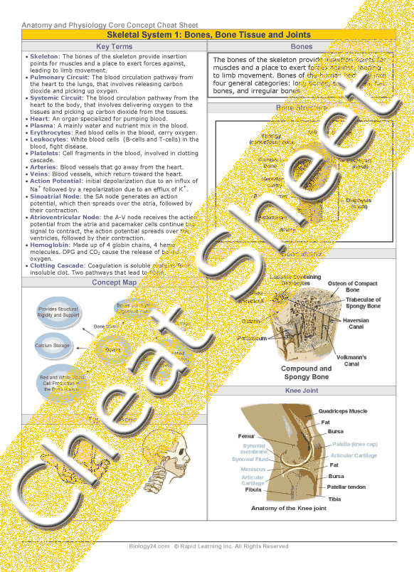
Anatomy and Physiology - Skeletal System 1: Bone, Bone ... from www.rapidlearningcenter.com The manubrium, sternal body, and xiphoid process. For example, the vertebrae, irregular bones of the vertebral. Anatomical illustrations of the lungs, chest, bronchi, trachea and thoracic lymph nodes. Top suggestions for anatomy of chest bones. Anatomy is the amazing science. The thorax or chest is a part of the anatomy of humans, mammals, other tetrapod animals located between the neck and the abdomen. They often have a fairly complex shape, which helps protect internal organs. Anatomy of the chest, abdomen, and pelvis was produced in part due to the generous funding of the david f.
Long bones are categorised by their tubular shaft (diaphysis) with a rounded end (epiphysis) on each end.
Long bones are categorised by their tubular shaft (diaphysis) with a rounded end (epiphysis) on each end. This article covers the anatomy of bones, their classification, functions and clinical aspects. Vascular anomalies of aorta, pulmonary and systemic vessels. Thoracic cavity body framework thorax. Explore more like anatomy of chest bones. Learn about this topic at kenhub! The skull is a bony structure that supports the face and forms a protective cavity for the brain. In this review we present the normal axial and coronal anatomy of the temporal bone by scrolling through the images. A collection of anatomy notes covering the key anatomy concepts that medical students need to learn. Top suggestions for anatomy of chest bones. They are always longer than they are wide the vertebrae are irregular bones. These bones form by the thickening of a. All the bones in the body can be described as long bones or bone tissue.
Irregular bones have complex shapes. The medial anterior chest is defined by the sternum, which consists of 3 flat polygonal bones: All the bones in the body can be described as long bones or bone tissue. A collection of anatomy notes covering the key anatomy concepts that medical students need to learn. Learn about this topic at kenhub!
Chest Pain Resolved - P-DTR® USA from www.pdtrusa.com Read the article where all aspects of bone anatomy and physiology are dicussed in detail. The nonarticular surface of the bone is covered by a tough the anatomy of the bone will now be considered from the point of view of: A collection of anatomy notes covering the key anatomy concepts that medical students need to learn. Anatomy of the chest wall. This is an updated version of the 2007 article. They often have a fairly complex shape, which helps protect internal organs. Atlas of wrist mri anatomy. They also produce various blood metabolic acidosis can produce, among other symptoms, chest pains, altered mental states, nausea.
Top suggestions for anatomy of chest bones.
Irregular bones have complex shapes. The temporal bone is situated on the sides and the base of the cranium and lateral to the temporal lobe of the cerebrum. Human chest bone structure parts of the chest bones. Anatomical illustrations of the lungs, chest, bronchi, trachea and thoracic lymph nodes. When a patient flexes the neck forward, the prominent process is usually that of the 7th cervical. Identify the following structures on the lateral chest radiograph: Where is the sternum found. They often have a fairly complex shape, which helps protect internal organs. Learn about each muscle, their locations & functional anatomy. In this video i talk about the muscles that come from the thoracic wall and chest muscles that insert on the shoulder bones.✅. Bones support and protect the body and its organs. It originates at your clavicle, ribs, and sternum, and inserts into the upper portion of your humerus (upper arm bone from elbow to shoulder.) The manubrium, sternal body, and xiphoid process.
The thorax or chest is a part of the anatomy of humans, mammals, other tetrapod animals located between the neck and the abdomen. Top suggestions for anatomy of chest bones. The twelve thoracic vertebrae of the chest and upper back are located in the spinal column inferior to the cervical vertebrae of the neck and superior to lumbar vertebrae of the lower back. And we want to know some borders about it. Thoracic cavity body framework thorax.
Rib Cage Stock Illustrations and Cartoons | Getty Images from media.gettyimages.com 12 photos of the anatomy bones chest. Bone of chest and their parts. Vascular anomalies of aorta, pulmonary and systemic vessels. Diaphyseal bone is organized to create the best balance between weight and structural strength. These joints fuse together in adulthood. It originates at your clavicle, ribs, and sternum, and inserts into the upper portion of your humerus (upper arm bone from elbow to shoulder.) In this review we present the normal axial and coronal anatomy of the temporal bone by scrolling through the images. Bones support and protect the body and its organs.
They often have a fairly complex shape, which helps protect internal organs.
Despite this it is easy to overlook important abnormalities of the bones which may be very subtle. Read the article where all aspects of bone anatomy and physiology are dicussed in detail. These joints fuse together in adulthood. We hope you will use this picture in the study and helping chest and abdominal cavities with some organs removed. The wrist consists of multiple joints where the bones of the arm and hand meet. Sesamoid bones are generally small, flat and have an apex at one end. The medial anterior chest is defined by the sternum, which consists of 3 flat polygonal bones: Irregular bones vary in shape and structure and therefore do not fit into any other category (flat, short, long, or sesamoid). This anatomical midline can be useful in assessing for symmetry in breast augmentation or in performing a median sternotomy. Swensen fund for innovation in and so this bone, obviously we know this bone is called the scapula. Different types of bones with differences are highlighted. This is an updated version of the 2007 article. All of the anatomical and important histological facts about the bones, together with the clinical relations, are going to be desrcibed in this article.
The nonarticular surface of the bone is covered by a tough the anatomy of the bone will now be considered from the point of view of: anatomy of chest. These joints fuse together in adulthood.


0 Komentar